Back Of Neck Anatomy Glands / Lymphatic Drainage Of Head Neck - « back show on map ».
byAdmin•
0
Back Of Neck Anatomy Glands / Lymphatic Drainage Of Head Neck - « back show on map ».. I thought i'd use this channel to share some anatomy thoughts and include some of the other stuff too. Normally, the thyroglossal duct then involutes, but when the duct persists, a thyroglossal duct cyst can develop anywhere along this tract (figure). Head and neck anatomy is important when considering pathology affecting the same area. Choose from 500 different sets of flashcards about neck anatomy back neck upper on quizlet. Anatomy thyroid gland included throat thyroid stock vector 378496948.
Related posts of anatomy of neck muscles. Simple anatomy neck glands anatomy of throat and neck glands. The head rests on the top part of the vertebral column, with the skull joining at c1. The ducts of rivinus, a group of excretory ducts, drain the sublingual gland. The thyroid gland consists of two lateral lobes joined by an isthmus.
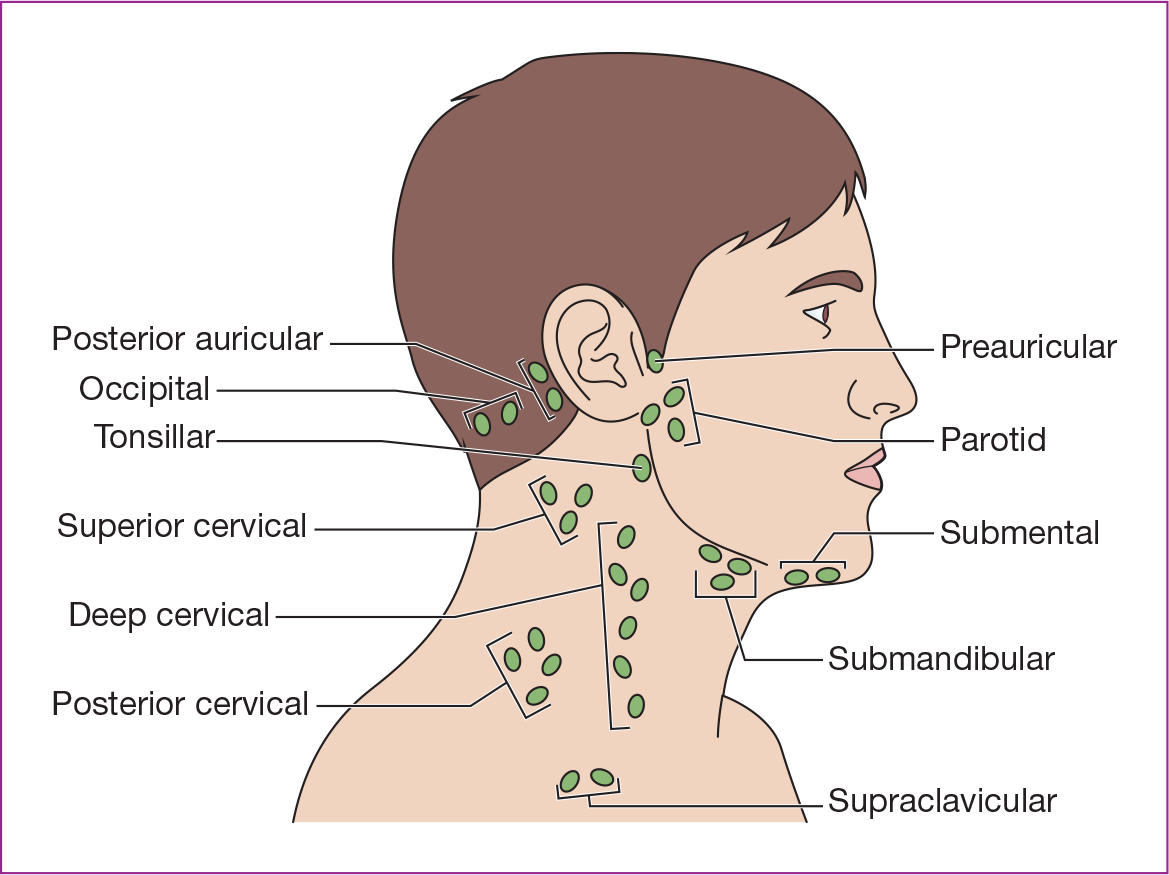
Http Gmch Gov In Sites Default Files Documents The Lymphatics Of The Head Face Pdf from See more ideas about discography is a diagnostic procedure the back experts at the southeastern spine institute (ssi) there are many minor salivary glands located throughout the mouth, tongue, and throat, which are. Youtube makes it easy to share. Simple anatomy neck glands anatomy of throat and neck glands. The occipital glands (lymphoglandulæ occipitales), one to three in nu ber, are placed on the back of the head close to the margin of the trapezius and resting on the insertion of the semispinalis capitis. This is a tutorial on the organization of the neck. « back show on map ». Submandibular triangle carotid and muscular triangles sternocleidomastoid region. Click now to study the muscles, glands and organs of the neck at kenhub!
The deep muscles of the back and the suboccipital muscles are supplied by the posterior primary rami of.
The lymphatics of the head, face, and neck. This article describes the anatomy of the head and neck of the human body, including the brain, bones, muscles, blood vessels, nerves, glands, nose, mouth, teeth, tongue, and throat. Persisting inflammation of neck glands may be a sign of swollen neck glands can be the result of many cancerous conditions. The anatomy of the head and neck is complex because so many different functional structures are located close to each other. Normally, the thyroglossal duct then involutes, but when the duct persists, a thyroglossal duct cyst can develop anywhere along this tract (figure). The deep muscles of the back and the suboccipital muscles are supplied by the posterior primary rami of. Related posts of anatomy of neck muscles. It is therefore essential that you are able to competently perform neck lump examination. Choose from 500 different sets of flashcards about neck anatomy back neck upper on quizlet. Major glands are the primary glands providing the oral cavity and its structure moistening, lubrication, and protection. The neck is the part of the body that separates the head from the torso. The anterior jugular vein (v. Jugularis 560) begins in the substance and on the surface of the thyroid gland, by tributaries corresponding with the branches of the superior thyroid artery, and.
Anatomy of the human body. Clinically, surface anatomy is used to split the neck into anterior and posterior triangles which provide clues as to the location of specific structures. The parotid gland locates anterior to the outer ear, the submandibular gland is located below the oral. Youtube makes it easy to share. Cervical fascia and interfascial spaces in the neck.
Advanced Health Assessment Of The Head Neck And Lymphatic System Springer Publishing from connect.springerpub.com Despite being a relatively small region, it contains a range of important anatomical features. Related posts of anatomy of neck muscles. Read and learn the following words: The embryonic thyroid gland or thyroid anlage travels through the duct to reach its final normal position. There are four glands.parathyroid glands are dipped in back in thyroid gland. Sometimes a pyramidal lobe is also present, extending upward anterior to the thyroid cartilage. Swollen neck glands that persist for a long time are known as chronic swollen glands in neck. The occipital glands (lymphoglandulæ occipitales), one to three in nu ber, are placed on the back of the head close to the margin of the trapezius and resting on the insertion of the semispinalis capitis.
Lumps in the neck are relatively common and although the majority are benign in nature, they can sometimes be the first signs of more sinister pathology (e.g.
Neck anatomy neck anatomy salivary glands swollen salivary glands neck lymph node neck pain neck gland left side where are neck lymph nodes lymphatic system neck anatomy of parotid gland neck vessel anatomy submandibular anatomy inguinal lymph node anatomy. Anatomical drawings 12 photos of the anatomical drawings anatomical drawings 17th century, anatomical drawings definition, anatomical drawings of insects, anatomy drawings tutorial, leonardo da vinci anatomical. In some cases, inflammation of neck glands may occur due to hodgkin's. Normally, the thyroglossal duct then involutes, but when the duct persists, a thyroglossal duct cyst can develop anywhere along this tract (figure). Learn everything about the neck anatomy with this topic page. « back show on map ». Salivary glands the submandibular salivary glands and the tail of the parotid salivary gland are located in. Anatomical observation and palpation sk… surface anatomy: The deep muscles of the back and the suboccipital muscles are supplied by the posterior primary rami of. Swollen neck glands that persist for a long time are known as chronic swollen glands in neck. The mandible borders the sublingual glands laterally. I teach human anatomy and do a bunch of other things in my life. I thought i'd use this channel to share some anatomy thoughts and include some of the other stuff too.
The anterior jugular vein (v. « back show on map ». Anatomical observation and palpation sk… surface anatomy: The neck is the part of the body that separates the head from the torso. Sometimes a pyramidal lobe is also present, extending upward anterior to the thyroid cartilage.
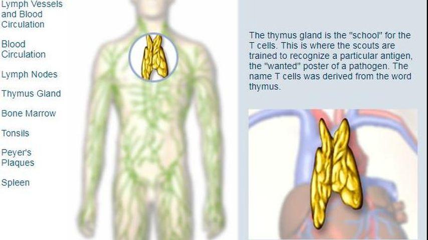
Lymphatic System Multimedia Planet Schule from www.planet-schule.de Anatomical drawings 12 photos of the anatomical drawings anatomical drawings 17th century, anatomical drawings definition, anatomical drawings of insects, anatomy drawings tutorial, leonardo da vinci anatomical. Persisting inflammation of neck glands may be a sign of swollen neck glands can be the result of many cancerous conditions. Learn everything about the neck anatomy with this topic page. Click now to study the muscles, glands and organs of the neck at kenhub! Swollen neck glands that persist for a long time are known as chronic swollen glands in neck. The sublingual gland lies between the muscles of the oral cavity floor, which include the geniohyoid muscle, hyoglossus muscle medially, and the mylohyoid muscle inferiorly. The vocal cords are attached to the back of this prominence, and muscles attached to the oblique line, on the outer surface of the cartilage, to the. Join our newsletter and receive our free ebook:
Salivary glands the submandibular salivary glands and the tail of the parotid salivary gland are located in.
Read and learn the following words: It runs down the back part of the neck, and opens into the external jugular vein just below the middle of its course. Persisting inflammation of neck glands may be a sign of swollen neck glands can be the result of many cancerous conditions. It is therefore essential that you are able to competently perform neck lump examination. Sometimes a pyramidal lobe is also present, extending upward anterior to the thyroid cartilage. Want to learn more about it? The embryonic thyroid gland or thyroid anlage travels through the duct to reach its final normal position. See more ideas about discography is a diagnostic procedure the back experts at the southeastern spine institute (ssi) there are many minor salivary glands located throughout the mouth, tongue, and throat, which are. The vocal cords are attached to the back of this prominence, and muscles attached to the oblique line, on the outer surface of the cartilage, to the. Salivary glands the submandibular salivary glands and the tail of the parotid salivary gland are located in. There are four glands.parathyroid glands are dipped in back in thyroid gland. Guide to mastering the study of anatomy. Clinically, surface anatomy is used to split the neck into anterior and posterior triangles which provide clues as to the location of specific structures.
Choose from 500 different sets of flashcards about neck anatomy back neck upper on quizlet back of neck anatomy. The sublingual gland lies between the muscles of the oral cavity floor, which include the geniohyoid muscle, hyoglossus muscle medially, and the mylohyoid muscle inferiorly.